Next-Generation Microfluidic Brain Implant Promises Revolutionary Precision in Neurological Research and Treatment

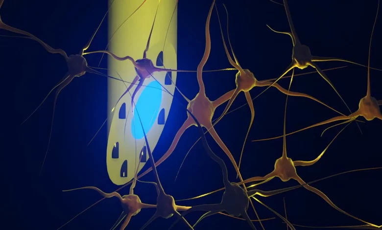
The landscape of neuroscience and neurological medicine may be on the brink of a profound transformation, courtesy of an international team of researchers who have engineered a groundbreaking neural interface. Recently detailed in the esteemed scientific journal Advanced Science, a newly developed brain implant known as the microfluidic Axialtrode, or mAxialtrode, introduces an unprecedented level of spatial precision for studying complex brain activity. By cleverly merging optical, electrical, and fluid-delivery capabilities into a single, highly flexible, needle-thin filament, this multi-institutional collaborative effort addresses some of the most persistent technological hurdles that have limited neuroscientific exploration for decades.
Conceived and developed through a partnership involving the Technical University of Denmark (DTU), the University of Copenhagen, and University College London (UCL), alongside other participating research bodies, the mAxialtrode represents a major leap forward from conventional brain-monitoring equipment. While the technology is presently optimized as an advanced research instrument to decode intricate neural pathways—such as those underlying epilepsy, cognitive processing, and memory—its long-term clinical potential is vast. Experts theorize that the device could eventually pave the way for closed-loop therapeutic interventions, whereby localized pharmaceuticals are administered simultaneously with targeted electrical or optical stimulation to treat debilitating neurological conditions.
The Genesis and Evolutionary Context of Brain-Computer Interfaces
To understand the magnitude of the mAxialtrode breakthrough, one must examine the evolutionary arc of brain implants and neuro-monitoring technologies. For decades, the neuroscientific community has relied on silicon-based microelectrodes and rigid metallic arrays to record electrical potentials from populations of neurons. While these traditional rigid implants have yielded foundational insights into brain function, they suffer from a critical biological incompatibility. The human brain is a soft, gelatinous organ that gently shifts within the skull, whereas traditional silicon and metal probes are stiff and unyielding. Over time, the micro-motions of the brain against a rigid foreign object cause mechanical irritation, localized tissue damage, and chronic neuroinflammatory responses. This immune reaction often results in glial scarring, which degrades the quality of the recorded electrical signals and eventually renders the implant ineffective.
Concurrently, researchers seeking to manipulate neural circuits turned to optogenetics—a revolutionary technique developed in the early 2000s that allows scientists to control genetically modified neurons with pulses of light. To deliver this light into deep brain structures, researchers typically inserted flat-ended optical fibers composed of glass or plastic. Although transformative, these conventional fibers presented their own geometric limitations. Light was emitted exclusively from the distal tip, or the very end of the fiber. Consequently, scientists could only stimulate or monitor a single focal point at a time. Because complex cognitive functions, sensory processing, and pathological states like epileptic seizures inherently rely on the synchronized communication across multiple distinct brain layers and deep subcortical structures, studying these phenomena required the insertion of multiple, cumbersome devices, compounding tissue trauma.
Addressing these historical limitations became the primary mission for Postdoc Kunyang Sui and Associate Professor Christos Markos at DTU, who spearheaded the design of the mAxialtrode. By rethinking the materials and architecture of the neural probe, the research team successfully integrated multiple functionalities into a single, tissue-friendly strand that minimizes physical damage while maximizing data collection and intervention capabilities.
Anatomy and Engineering of the mAxialtrode
The structural composition of the mAxialtrode distinguishes it starkly from its predecessors. The device begins its lifecycle as a macro-scale polymer rod. Utilizing a specialized thermal drawing process akin to fabricating a fine strand of glass or sugar under tightly controlled tension, researchers scale down the polymer rod into an extremely thin, flexible fiber measuring less than half a millimeter across.
At the structural core of this polymer filament lies a dedicated light-conducting pathway designed to transmit optical signals efficiently across deep tissue regions. Encircling this central optical core are eight microscopic microfluidic channels. These minuscule fluid conduits serve a dual purpose: they are capable of transporting minute, precisely measured volumes of liquid drugs or chemical agents to targeted cellular environments, and they can house ultra-thin metallic micro-wires. These embedded wires function as high-resolution electrical sensors capable of detecting the faint ionic currents generated by firing neurons.
Because the entire apparatus is constructed from soft, flexible, polymer-based materials rather than brittle silicon or hard metals, the mAxialtrode mimics the mechanical compliance of native brain tissue. When implanted, the fiber flexes harmoniously with the natural micro-movements of the brain, drastically reducing chronic friction and mitigating the inflammatory immune responses that plague traditional hard probes. Furthermore, the device features a specially engineered angled tip. This geometric optimization eases the insertion trajectory, further minimizing the initial trauma inflicted upon delicate neural architecture during the implantation procedure.
Rigorous Validation in In Vivo Models
Moving from theoretical design to empirical validation required stringent testing in living biological systems. The DTU research team partnered closely with neurophysiology experts, including Associate Professor Rune W. Berg from the University of Copenhagen and Associate Professor Rob C. Wykes from University College London, both of whom brought deep expertise in neural circuit analysis and translational epilepsy models.
The device was subjected to comprehensive in vivo testing within living murine models. During these experiments, the needle-thin mAxialtrode was surgically implanted into the brains of the test subjects and coupled to external hardware, including miniature fluid pumps, light sources, and sophisticated electrophysiological recording equipment.
The experimental outcomes exceeded expectations. The mAxialtrode successfully demonstrated the capacity to stimulate targeted nerve cells using dual-wavelength capabilities—specifically employing blue and red light to activate light-sensitive neuronal populations. Simultaneously, the embedded micro-wires recorded clean, high-fidelity electrical activity across both shallow and deep anatomical structures, including the cerebral cortex and the hippocampus.
Perhaps most impressively, the researchers utilized the internal microfluidic channels to inject distinct chemical substances at varying depths within the brain during real-time monitoring. The delivery points were spaced nearly three millimeters apart along a single implant trajectory, enabling localized pharmacological intervention while concurrently recording the immediate electrophysiological response. Throughout these complex procedures, the test subjects carried the lightweight, integrated fiber array without exhibiting any visible signs of behavioral distress or physical discomfort.
Implications for Epilepsy Research and Clinical Neurology
The successful validation of the mAxialtrode opens expansive new avenues for neuroscientific inquiry, with immediate implications for the study of epilepsy—a chronic neurological disorder characterized by recurrent, unprovoked seizures that affect tens of millions of individuals worldwide.
Understanding how epileptic seizures originate, propagate through distinct cortical layers, and terminate has historically been hindered by the inability to simultaneously record, stimulate, and pharmacologically modulate interconnected brain regions in real time. The mAxialtrode offers a consolidated solution. By allowing researchers to monitor electrical discharges while instantly suppressing aberrant neural firing via targeted drug delivery or localized optical stimulation, the device provides a powerful platform for dissecting the pathophysiology of epilepsy.
Associate Professor Rob C. Wykes of UCL emphasized the clinical relevance of these capabilities, noting that the ability to deliver anti-epileptic compounds directly to an epileptic focus while tracking real-time electrical activity could fundamentally change how focal drug delivery systems are developed. In traditional pharmacological treatments, systemic administration of anti-seizure medications often results in severe systemic side effects because the drugs circulate throughout the entire body rather than concentrating solely on the hyper-excitable neural tissue. A localized, responsive delivery system could theoretically maximize therapeutic efficacy while minimizing adverse side effects.
Chronology of Development and Collaborative Framework
The path leading to the publication in Advanced Science represents years of cross-disciplinary collaboration among top-tier European research institutions:
- Conceptualization Phase: DTU researchers Kunyang Sui and Christos Markos conceptualize a unified platform capable of combining optical transmission, electrical recording, and fluidics into a single soft fiber.
- Material Engineering and Fabrication: The team optimizes the thermal drawing process, successfully scaling polymer rods down to sub-millimeter flexible fibers equipped with microscopic internal channels.
- Interdisciplinary Partnerships: DTU joins forces with the University of Copenhagen and University College London, integrating expertise in polymer physics, neurophysiology, and clinical epilepsy models.
- In Vivo Validation: Preclinical trials are conducted in living subjects, confirming the device’s efficacy in simultaneous optical stimulation, electrical recording, and multi-depth fluid delivery.
- Publication and Patenting: Findings are peer-reviewed and published in Advanced Science, while the research consortium initiates patent filings to protect the intellectual property surrounding the mAxialtrode architecture.
Roadmap to Clinical Translation and Future Outlook
Despite the enthusiasm generated by the publication of the mAxialtrode, the scientific team maintains a measured and cautious perspective regarding its immediate clinical availability. Postdoc Kunyang Sui has explicitly stressed that while the technology represents a monumental breakthrough for laboratory research, it remains in the pre-clinical stage and is still a considerable distance from routine human clinical use.
Translating any implantable neural device from animal models to human patients requires a rigorous, multi-step pipeline. The development roadmap ahead includes extended biocompatibility studies to assess the long-term safety of the polymer materials over months and years of continuous implantation, scaling manufacturing processes to ensure absolute reproducibility, and navigating stringent regulatory pathways overseen by medical device authorities such as the European Medicines Agency (EMA) and the U.S. Food and Drug Administration (FDA).
Nevertheless, the foundational architecture of the mAxialtrode provides a versatile template for future generations of neuro-technologies. As researchers continue to refine the device, expand the number of internal microfluidic channels, and explore wireless integration for fully implantable systems, the dream of smart, closed-loop neuro-therapeutics moves steadily closer to reality.
For the broader scientific community, the mAxialtrode stands as a testament to the power of convergent engineering—where materials science, optics, fluid dynamics, and neurobiology intersect to push the boundaries of what is technologically possible in the exploration of the human brain.