Decoding the Gut: Johns Hopkins Researchers Discover How a Common Bacterium Triggers Colon Cancer and Design a Molecular Decoy to Block It

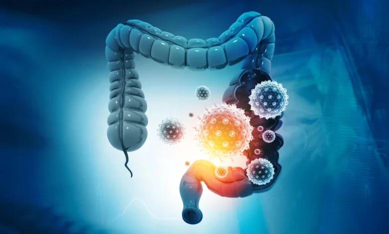
For nearly two decades, the scientific community has grappled with an unsettling biological paradox: a common bacterium residing quietly in the intestines of up to 20 percent of healthy people can, under specific conditions, produce a potent toxin capable of driving tumor formation in the colon. While researchers long ago established the epidemiological link between this microbe and colorectal malignancies, a foundational mystery persisted. Scientists understood that the toxin wreaked havoc once inside the tissue, but they remained entirely in the dark regarding how the molecule first attached itself to intestinal cells to initiate its assault.
That missing link has finally been uncovered. In a breakthrough published in the journal Nature, an international team led by researchers at the Johns Hopkins Kimmel Cancer Center, the Bloomberg-Kimmel Institute for Cancer Immunotherapy, and the Johns Hopkins University School of Medicine has identified the exact molecular docking point utilized by the bacterial toxin. This discovery not only solves a long-standing puzzle in gastrointestinal microbiology but has already allowed scientists to engineer a decoy molecule that successfully intercepts the toxin in animal models, blazing a potential new trail for the prevention of bacterial-driven colorectal cancer.
Chronology of a Two-Decade Scientific Pursuit
The journey toward this recent discovery spans nearly twenty years of methodical oncological and microbiological research, marked by critical milestones that gradually illuminated the connection between gut flora and cancer.
The foundational piece of this puzzle was placed in 2009, when senior author Cynthia Sears, M.D., alongside her colleagues, published a landmark study in Nature Medicine. That research provided definitive evidence that a specific toxin-producing strain of the common gut bacterium Bacteroides fragilis could actively promote the development of colon tumors in laboratory models. The findings helped cement a paradigm shift in oncology, proving that microbes could play a direct, active role in carcinogenesis through chronic inflammation and tissue disruption.
In the years following the 2009 study, Sears and other research teams dove deeper into the mechanism of action. They discovered that the culprit substance, known as B. fragilis toxin (or BFT), targets a specific structural protein called E-cadherin. E-cadherin acts as a cellular mortar, holding neighboring epithelial cells together and maintaining the integrity of the colon’s protective mucosal barrier. When BFT cleaves E-cadherin, the barrier fractures. This compromise allows luminal contents, bacteria, and inflammatory agents to seep into surrounding tissues, triggering a cascade of immune responses and cellular changes favorable to tumor growth.
However, a frustrating roadblock continually stymied the investigators. Despite exhaustive biochemical analyses, experiments revealed that BFT did not bind directly to E-cadherin. The toxin clearly required an intermediary or an initial docking site to get a foothold on the cell surface before it could reach and dismantle E-cadherin. For over a decade, attempts to identify this missing receptor repeatedly failed, leaving a critical gap in the understanding of the pathogenic mechanism.
Leveraging CRISPR Technology to Find the Missing Receptor
To break through this multi-decade impasse, the Johns Hopkins team changed tactics, turning to advanced genetic engineering. Maxwell White, an M.D./Ph.D. candidate in the Sears laboratory, spearheaded a genome-wide CRISPR screening initiative in close collaboration with the laboratory of Matthew Waldor at Harvard Medical School.
CRISPR technology allows scientists to systematically disable individual genes within a living cell to observe the functional consequences. By applying this high-throughput genetic screen to human colon epithelial cells—the primary cells forming the inner lining of the colon—the research team sought to pinpoint precisely which genes were mandatory for BFT to successfully latch onto and damage the cells.
Out of thousands of candidates, one gene emerged as a resounding top hit. It was the gene responsible for producing claudin-4, a well-known protein localized in tight junctions. Tight junctions are the intercellular seals that regulate paracellular permeability, ensuring that substances cannot freely cross the intestinal barrier without regulation.
When the researchers selectively disabled the claudin-4 gene in the laboratory cultures, the results were instantaneous and definitive: BFT could no longer bind to the colon cells. Deprived of its primary anchoring point, the toxin was completely rendered unable to reach or cleave its secondary target, E-cadherin. Claudin-4 was thus unmasked as the essential cell-surface receptor required for the bacterium’s toxic campaign.
A Surprising Twist in Toxin Biology
The identification of claudin-4 as the BFT receptor stunned the scientific team. Prior to the CRISPR screen, researchers had strongly hypothesized that BFT—acting as a protease (an enzyme that cuts other proteins)—would dock via a signaling receptor, such as a G protein-coupled receptor (GPCR). GPCRs are classic communication switches embedded in cell membranes that allow cells to interpret and respond to external chemical signals.
Instead, the toxin hijacked a structural component designed to maintain tissue architecture. This realization also illuminated an unusual structural and functional pathway. While most proteases interact directly with their final targets, BFT requires a distinct, non-signaling structural protein just to position itself for the subsequent destruction of E-cadherin. A comprehensive review of existing literature confirmed that no other known bacterial toxin utilizes this exact two-step mechanism.
To validate the physical interaction between the two molecules, the Johns Hopkins team partnered with structural biologists F. Xavier Gomis-Rüth and Ulrich Eckhard at the Molecular Biology Institute of Barcelona. Using state-of-the-art biophysical assays designed to measure molecular binding affinities, the researchers demonstrated that BFT and claudin-4 form an exceptionally tight complex, pairing up on a strict one-to-one molecular ratio. This provided undeniable physical evidence of the docking mechanism.
Translating the Discovery Into a Molecular Decoy
Pinpointing the receptor did more than satisfy a theoretical curiosity; it provided an immediate blueprint for therapeutic intervention. Reasoning that if BFT requires claudin-4 to bind to colon cells, a synthetic substitute could theoretically lure the toxin away, the research team designed a molecular decoy.
This decoy consisted of a soluble protein engineered to carry the specific binding sequences of claudin-4. The objective was to create a decoy target that would circulate freely, intercepting BFT in the intestinal environment and neutralizing it before it ever made contact with the actual cellular lining of the colon.
To test this hypothesis in a complex biological setting, the Johns Hopkins group teamed up with the laboratory of Min Dong at Harvard Medical School, including researcher Kang Wang and his colleagues. The decoy strategy was tested in murine models exposed to the bacterial toxin.
The experimental results exceeded expectations. In the mice treated with the molecular decoy, BFT preferentially bound to the soluble decoy proteins rather than the claudin-4 receptors on the colon epithelial cells. Consequently, the decoy successfully intercepted the toxin and completely protected the murine subjects from the tissue-damaging effects characteristic of BFT exposure.
Broader Implications and Future Horizons
The successful deployment of a molecular decoy in animal models marks a significant conceptual shift in how scientists might approach toxin-mediated diseases. Rather than attempting to repair the extensive tissue damage or inflammation after it has already occurred, future therapeutics could intercept the destructive process at its absolute inception.
Senior author Cynthia Sears emphasized the broad clinical potential of the discovery. Understanding the precise biochemical pathways of bacterial toxins, she noted, opens new avenues not just for oncology, but for the diagnosis and treatment of a spectrum of associated conditions, ranging from persistent diarrhea to severe systemic bloodstream infections.
Despite the enthusiasm surrounding the breakthrough, researchers urge caution and outline a rigorous path ahead. While the decoy strategy successfully shielded mice from acute tissue damage, the current experiments did not explicitly prove that the decoy prevents the long-term development of colorectal cancer. Furthermore, researchers have yet to capture the exact high-resolution three-dimensional crystal structure of the BFT-claudin-4 complex. Even cutting-edge artificial intelligence platforms, such as AlphaFold, have thus far been unable to fully resolve the intricate geometry of the molecular interface. Acquiring this structural map will be crucial for the rational design of optimized pharmaceutical compounds.
Moving forward, Maxwell White and the research team are actively investigating how to refine the decoy approach into viable pharmacological treatments. This includes exploring both small-molecule compounds and biologic drugs that boast superior pharmacokinetic properties, stability, and administration profiles in the human body.
As investigations continue, this foundational discovery successfully bridges a twenty-year knowledge gap in cancer microbiology. By revealing the microscopic handshake that allows a common gut bacterium to initiate its carcinogenic attack, scientists have provided a concrete target—and a promising defense mechanism—in the ongoing effort to mitigate the burden of colorectal cancer.