
Unlocking the Blueprint of Movement: Researchers Discover Universal Tendon and Ligament Stem Cells With Implications for Spinal Stenosis

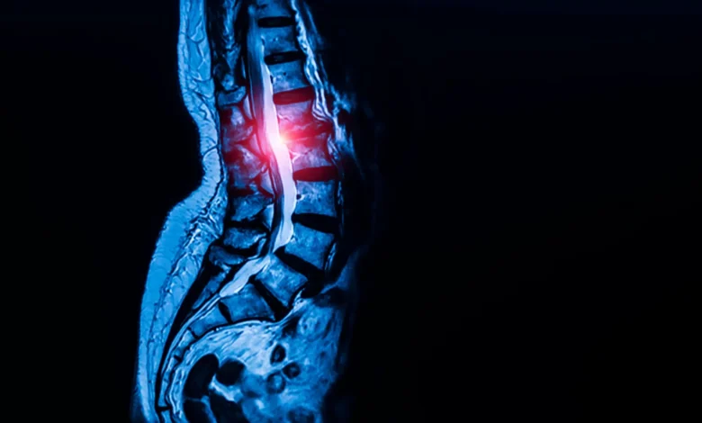
In a monumental breakthrough for musculoskeletal medicine, a collaborative team of researchers at Weill Cornell Medicine and the Hospital for Special Surgery (HSS) has successfully identified a previously elusive, universal population of stem cells responsible for generating the body’s tendons and ligaments. Published in the peer-reviewed journal Cell on September 7, the discovery closes a long-standing knowledge gap in regenerative biology. More than just a foundational scientific milestone, the identification of these specialized cells has immediate translational implications, offering a fresh mechanistic perspective on lumbar spinal stenosis—a debilitating degenerative condition affecting more than 100 million people worldwide.
By isolating the elusive progenitor cells and tracing their activity, the research team not only mapped out the cellular architecture governing connective tissues throughout the human body but also pinpointed a hyperactive signaling pathway driving spinal stenosis. Crucially, this biological insight points toward an unexpected therapeutic avenue: existing FDA-approved hypertension medications, known as calcium channel blockers, could potentially be repurposed to halt or reverse the progression of spinal narrowing without invasive surgery.
The Anatomy of the Discovery and the Challenge of "Stemness"
For decades, developmental biologists and orthopedic researchers have understood the broad mechanics of skeletal and muscular systems while remaining largely in the dark about the exact lineage of connective tissues. While scientists had previously mapped out skeletal stem cells—including pioneering work by the same Weill Cornell team in 2018 that identified bone-repair and skull-forming stem cells—finding an equivalent progenitor population for tendons and ligaments proved exceptionally difficult.
The core obstacle lay in cellular camouflage. Unlike bone tissue, which possesses distinct structural markers, tendons and ligaments are densely populated with fibroblast-like cells that appear morphologically identical under standard microscopic examination. Differentiating a mature, static fibroblast from an active, self-renewing stem cell was akin to finding a single specific grain of sand on a vast beach.
To overcome this hurdle, the research team—led by co-corresponding authors Dr. Matthew Greenblatt, associate professor of pathology and laboratory medicine at Weill Cornell, and Dr. Sravisht Iyer, associate professor of orthopedics at Weill Cornell and spine surgeon at HSS—leveraged advanced single-cell genomic technologies. Working alongside first author Dr. Lingling Hu, a postdoctoral fellow, the team analyzed thousands of individual cells harvested from murine models, sorting them by genetic expression profiles to identify which populations exhibited the definitive properties of "stemness."
In stem cell biology, "stemness" is defined by a dual capability: the capacity for indefinite self-renewal coupled with the multipotent ability to differentiate into the full spectrum of mature cell types required to build and maintain a specific tissue. The researchers discovered these rare cells residing within specialized niches inside tendons and ligaments, functioning as a biological reservoir designated for tissue growth and repair.
Validation in Human Tissues and Ubiquitous Distribution
Following the successful isolation of these cells in mice, the researchers transitioned to human tissues to confirm translational relevance. Human ligament samples were procured with strict informed consent protocols from patients undergoing necessary spinal decompression surgeries performed by Dr. Iyer.
Through rigorous cellular assays, Dr. Hu and the research team demonstrated that these human-derived cells maintained the same dual characteristics observed in animal models: they could both proliferate to maintain their own numbers and successfully differentiate into functional ligament cells. Furthermore, when the researchers mapped the anatomical distribution of these progenitor cells, they discovered they were not restricted to the spinal column.
Subsequent histological and cellular analyses of human tissue samples revealed the presence of these exact stem cells across diverse anatomical locations, including the patellar ligament of the kneecap and the Achilles tendon.
"We looked in the kneecap ligament; we looked at the Achilles tendon; and everywhere we looked, we found this cell," Dr. Greenblatt noted. "So, we think this is the universal stem cell for tendons and ligaments throughout the body."
Unraveling the Pathogenesis of Lumbar Spinal Stenosis
With the universal stem cell identified, the investigative team turned their attention to clinical pathology, focusing specifically on lumbar spinal stenosis. This degenerative condition develops when spinal ligaments thicken over time, severely narrowing the spinal canal. The resulting compression on adjacent neural pathways manifests as chronic lower back pain, numbness, radiating discomfort, and profound difficulty walking or maintaining mobility. Historically, treatment options have been limited to conservative physical therapy or major surgical decompression once the condition reaches an advanced, debilitating stage.
To determine whether the newly discovered stem cells played a direct role in this disease process, the researchers conducted comparative phenotypic analyses. They examined stem cells harvested from patients suffering from severe lumbar spinal stenosis and contrasted them with control samples collected from spinal ligaments of patients undergoing surgery for herniated discs, who showed no clinical signs of stenosis.
The findings were striking. Ligaments derived from patients with spinal stenosis harbored a significantly higher concentration of these stem cells compared to control tissues. Furthermore, when these stenosis-derived stem cells were transplanted into experimental mouse models, they exhibited a marked propensity for hyper-proliferation, generating excessive tendon and ligament tissue far beyond normal homeostatic requirements.
"Though spinal stenosis is a complex condition, this really showed us that these cells are contributing to the pathology," Dr. Greenblatt explained, emphasizing that the disease is driven, at least in part, by an overactive cellular engine rather than simple mechanical wear and tear.
Intracellular Signaling and the Calcium Connection
Delving deeper into the cellular machinery, the researchers sought to uncover the exact molecular triggers causing these stem cells to run amok. Their investigations revealed that stem cells isolated from spinal stenosis patients displayed significantly elevated intracellular calcium signaling compared to their healthy counterparts.
Calcium signaling serves as a vital biochemical language within cells, governing myriad physiological processes, including proliferation, metabolic activity, and structural growth. To test whether this elevated signaling was a mere correlate or a direct causal mechanism, the researchers engineered experiments to artificially increase calcium signaling within healthy stem cells. The result was rapid, abnormal tissue expansion mirroring the pathology of spinal stenosis.
Conversely, when the research team therapeutically dampened calcium signaling within a murine model of lumbar spinal stenosis, the pathological tissue growth was successfully halted and blocked. This pivotal discovery transformed a complex degenerative condition into a tractable pharmacological target.
A New Horizon in Pharmacological Interventions
The identification of calcium signaling pathways as the primary driver of pathological ligament thickening opens an unprecedented door in orthopedic pharmacology. Because the mechanisms governing this abnormal cell growth are tied to cellular calcium regulation, the research team immediately recognized a potential bridge to existing pharmacopeia: calcium channel blockers.
Calcium channel blockers are a well-established, widely prescribed class of medications routinely used to manage systemic hypertension and various cardiovascular conditions. Because their safety profiles, pharmacokinetic properties, and side-effect parameters are already extensively documented through decades of clinical use across millions of patients, the prospect of drug repurposing offers a streamlined path toward clinical trials.
While researchers caution that rigorous, controlled clinical trials will be mandatory to determine both the safety and efficacy of administering calcium channel blockers locally or systemically for spinal stenosis, the clinical implications are profound.
"This is probably the first work that’s shown a potential therapeutic target for one of the most common spine conditions in the world," Dr. Iyer remarked. "Identifying these specialized stem cells unlocks a new area of research that allows us to address this disease much more mechanistically, rather than just waiting until a patient’s condition worsens and requires surgery to relieve the nerve compression. The findings are exciting for their potential to change the way we deliver spinal care."
Broader Implications for Connective Tissue Disorders and Regenerative Medicine
Beyond lumbar spinal stenosis, the mapping of this universal tendon and ligament stem cell holds transformative potential for a broad array of musculoskeletal conditions that have long frustrated clinicians. Tendons and ligaments possess notoriously poor intrinsic healing capabilities due to a lack of robust vascular supply and low cellular turnover, making injuries such as rotator cuff tears, severe Achilles tendon ruptures, and chronic tendinopathies notoriously difficult to manage and prone to re-injury.
Dr. Greenblatt and his colleagues plan to expand their research initiatives to investigate whether functional defects or signaling irregularities within this universal stem cell population contribute to systemic connective tissue disorders, such as Marfan syndrome—a genetic condition that profoundly impacts the body’s structural integrity. By understanding how these stem cells regulate matrix production in both healthy and diseased states, regenerative medicine researchers may eventually design targeted biologic therapies to stimulate natural tissue repair, potentially revolutionizing orthopedic surgery and sports medicine alike.
The foundational research was made possible through extensive collaborative funding and institutional support, backed by grants and fellowships from organizations including the National Institutes of Health, the Marfan Foundation, the Arthritis National Research Foundation, the Children’s Tumor Foundation, the Mary Kay Ash Foundation, the Pershing Square Foundation, and Weill Cornell’s internal research initiatives. As these findings transition from the laboratory bench toward future clinical evaluations, millions of patients suffering from debilitating degenerative connective tissue diseases may finally see a horizon defined by pharmacological innovation rather than surgical inevitability.