How Disrupted Deep Sleep and Alzheimer’s Protein Accumulation Undermine Memory in the Aging Brain

Every day, millions of people experience a momentary, frustrating lapse in memory: walking out of a grocery store and struggling to recall where the vehicle was parked, or forgetting the name of a familiar
acquaintance met in passing. While these minor cognitive hiccups are frequently dismissed as a normal byproduct of getting older, neuroscientists have long sought to understand the precise neurological mechanisms driving this decline. A groundbreaking study recently published in the prestigious journal Nature Neuroscience sheds new light on this phenomenon, offering compelling evidence that connects microscopic changes in the aging brain to the subtle fraying of episodic memory.
Led by researchers at the University of California, Berkeley, the investigation points to an intersection between declining memory performance, disrupted electrical activity during deep sleep, and the accumulation of tau—a hallmark protein associated with Alzheimer’s disease. Spearheaded by neuroscientist Omer Sharon and conducted under the direction of former Berkeley sleep researcher Matthew Walker, the research team discovered that the buildup of tau in the brain’s frontal cortex directly interferes with the synchronized electrical waves that help consolidate memories overnight. These findings not only illuminate the complex relationship between sleep architecture and cognitive health but also provide a new lens through which scientists can view the earliest, preclinical stages of neurodegeneration.
The Anatomy of Episodic Memory and the Aging Brain
At the heart of human recollection is episodic memory, the cognitive system responsible for encoding, storing, and retrieving autobiographical events from our lives. From monumental milestones, such as a child’s first day of elementary school, to mundane daily occurrences like remembering where one left a set of keys, episodic memories form the foundation of our long-term personal narrative. Unfortunately, this foundational cognitive faculty is among the first to falter as individuals age.
As decades pass, the rich sensory details—the sights, sounds, and contextual cues—associated with past experiences become increasingly difficult to retrieve. While some degree of age-related cognitive slowing is considered a standard component of human maturation, these changes can escalate dramatically in individuals developing Alzheimer’s disease or other forms of dementia. For decades, the exact physiological bridge between normal age-related memory loss and the pathological changes of Alzheimer’s remained elusive. The UC Berkeley study bridges this gap by demonstrating that microscopic protein pathology can disrupt normal brain function long before clinical symptoms of cognitive impairment manifest.
The Critical Role of Deep Sleep in Memory Consolidation
To understand how tau protein disrupts memory, researchers must first examine the mechanics of human sleep. Sleep is not a uniform state of rest; rather, it is a dynamic cycle composed of distinct stages, each characterized by unique patterns of electrical brain activity. While rapid eye movement (REM) sleep is widely recognized for hosting vivid dreams and facilitating emotional processing, non-REM (NREM) sleep plays an indispensable role in maintaining fundamental brain health and cognitive resilience.
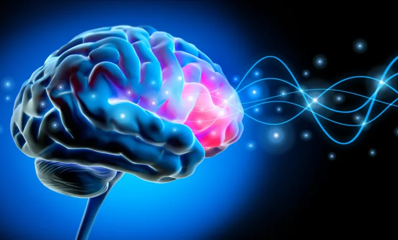
During the deepest phases of NREM sleep—a period largely devoid of dreaming—large populations of neurons undergo a synchronized physiological choreography. Millions of brain cells briefly switch off and back on together in a highly coordinated sequence. These mass neuronal transitions generate massive, slow electrical waves that ripple across vast expanses of the brain, typically originating in the frontal cortex and cascading outward.
"Each slow wave reflects an enormous population of neurons switching off and back on together," explained Omer Sharon, a postdoctoral researcher at UC Berkeley’s Center for Human Sleep Science. "Our study shows that for memory, it’s critical that these events cascade in sequence across large parts of the brain. This coordinated shutdown has been observed only during deep sleep, and may be one reason why we sleep."
The Breakdown of Synchronized Neural Activity
When the brain is functioning optimally, these traveling slow waves act as a systemic courier service, transferring newly acquired information from temporary holding areas in the hippocampus to the more permanent storage vaults of the neocortex. However, the UC Berkeley research reveals that this delicate orchestration begins to fail as tau protein accumulates within the aging brain.
Using high-density electroencephalograms (EEGs) to monitor sleeping brain activity, the researchers compared a cohort of cognitively healthy young adults in their early 20s with an older demographic ranging in age from their mid-60s to mid-70s. The comparative data revealed stark contrasts. In young participants, slow-wave discharges moved cohesively across the scalp in broad, sweeping patterns roughly equivalent to the width of a hand. In contrast, the older participants exhibited fragmented, isolated electrical activity. Instead of traveling together in harmonious waves, the electrical impulses appeared sporadic, covering shorter distances before fading away.
The research team coined a poignant descriptor for these fractured electrical signals: "lonely waves." To test the functional consequences of these disconnected patterns, the investigators subjected participants to memory retention trials. Before going to sleep, participants were asked to memorize pairs of associated words. The following morning, their ability to recall these pairings was tested to measure overnight memory consolidation.
The results established a direct correlation. Participants whose brains produced more "lonely waves" during deep sleep consistently demonstrated inferior overnight memory retention. Furthermore, tracking a subset of these older adults over a multi-year longitudinal period revealed that as tau levels in the frontal cortex increased, slow waves grew progressively less coordinated, leading to a measurable decline in the ability to retain newly learned information.
Connecting Sleep Disruption to Alzheimer’s Pathology
To confirm that these aberrant sleep patterns were tied directly to Alzheimer’s-related pathology rather than general aging alone, the research team enlisted the expertise of Dr. William Jagust, a UC Berkeley professor emeritus of public health and neuroscience. Utilizing positron emission tomography (PET) scans, the researchers mapped the distribution of tau protein within the brains of the older participants by introducing safe, radioactive tracers into the bloodstream.
The PET imaging confirmed that greater accumulations of tau in the frontal cortex directly corresponded to the breakdown of long-distance traveling slow waves during deep sleep. Crucially, the participants exhibiting these neurological signatures had not developed Alzheimer’s disease.
"These people did not have Alzheimer’s," Sharon emphasized. "They had tau in their brains, but with subclinical impacts. Their memory decline was within the normal range for their age."
This distinction is vital for understanding the trajectory of neurodegenerative research. The findings suggest that the microscopic accumulation of tau operates on a continuum, influencing cognitive performance and sleep architecture long before a clinical diagnosis of dementia is warranted.
Corroborating Findings Through Cerebrospinal Fluid Analysis
Because PET imaging facilities are costly and require specialized medical infrastructure, the research team sought to validate their findings through an alternative methodology. They collaborated with neurologist Dr. Yo-El Ju at Washington University in St. Louis to analyze cerebrospinal fluid (CSF) samples obtained from a distinct, independent cohort of older adults.
While spinal fluid analysis cannot pinpoint the precise anatomical location of tau accumulation within the frontal cortex in the same manner as a PET scan, the biochemical measurements corroborated the imaging data. Individuals whose sleeping brains generated higher frequencies of isolated slow waves exhibited elevated ratios of tau to amyloid-beta—another primary protein aggregate implicated in the pathogenesis of Alzheimer’s disease.
The convergence of data from multiple independent cohorts, utilizing distinct technological modalities including EEGs, PET scans, and CSF assays, significantly strengthened the credibility of the conclusions. "It’s collected in a totally different place and in a different cohort, so I think that adds to the strength of the evidence," Sharon noted.
Broader Implications for Future Diagnostics and Therapeutics
The implications of this study extend far beyond theoretical neuroscience, offering potential pathways for future clinical interventions. By establishing that cognitive decline in aging is not merely an immutable consequence of the passage of time, but rather tied to specific, measurable physiological disruptions, the research opens new avenues for therapeutic development.
"It’s not just about age," Sharon remarked. "It’s how much pathology you have in frontal areas where global waves originate."
As researchers look toward the future, the primary challenge will be establishing causality within this complex pathophysiological triad. While the longitudinal data clearly demonstrates that tau accumulation and lonely waves increase in tandem over time, scientists have yet to definitively prove whether accumulating tau directly causes the breakdown of synchronized sleep waves, whether sleep fragmentation accelerates tau deposition, or if an underlying third factor drives both phenomena.
"We can see tau and lonely waves rising together over time," Sharon concluded. "What we cannot yet say is which leads—and that question will shape how we design interventions."
Ultimately, the study underscores the profound importance of deep sleep not just as a period of physical restoration, but as a critical window during which the brain actively maintains its cognitive integrity. By monitoring the architecture of deep sleep, future medical professionals may gain an invaluable early-warning system for detecting neurodegenerative pathology, allowing for timely therapeutic interventions designed to preserve memory and cognitive health across the human lifespan.