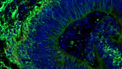
Beyond Amyloid and Tau: Researchers Uncover 3D Genome Disruption as a Novel Driver of Alzheimer’s Disease


A collaborative team of researchers spanning Carnegie Mellon University, the University of Pittsburgh School of Medicine, and the University of Washington has unveiled a groundbreaking perspective on the pathology of Alzheimer’s disease. Published in the prestigious journal Science, the study shifts the scientific gaze away from the traditional, singular focus on amyloid-beta plaques and tau tangles, directing attention instead toward the three-dimensional architecture of the genome within individual brain cells. By demonstrating how the physical folding of DNA is fundamentally altered in the brains of Alzheimer’s patients, this research opens critical new frontiers for therapeutic intervention, disease modeling, and early detection.
The Main Facts: Unlocking the Three-Dimensional Genome
For decades, the global scientific consensus surrounding Alzheimer’s disease has been anchored to two principal molecular hallmarks: the extracellular accumulation of amyloid-beta plaques and the intracellular aggregation of hyperphosphorylated tau proteins. While these hallmarks remain foundational to understanding neurodegeneration, therapies targeting them have yielded mixed clinical results, slowing cognitive decline in some patients but failing to halt or reverse the disease entirely. This limitation has driven investigators to search for deeper, more systemic layers of cellular dysfunction.
The newly published study reveals that the three-dimensional (3D) folding of the genome in specific brain cell types differs significantly between healthy individuals and those suffering from Alzheimer’s disease. DNA is not housed within a cell nucleus as a linear, uncoiled string; rather, it is meticulously folded into complex 3D structures that dictate whether specific genes remain accessible and active. When this physical organization is compromised, the cell’s regulatory framework breaks down.
Using an innovative confluence of single-cell multi-omics, spatial transcriptomic mapping, and a bespoke deep learning framework named Hicformer, the research team successfully mapped how genome folding correlates directly with gene activity and tissue-level degradation within the prefrontal cortex—a critical region of the brain responsible for executive function, decision-making, and complex cognitive behaviors. The study establishes that higher-order chromatin alterations are not merely incidental phenomena, but integral components of the molecular pathology driving Alzheimer’s.
Historical Context and Chronology of the Discovery
To appreciate the significance of this breakthrough, one must contextualize the evolution of genomic research in neurodegenerative diseases. Historically, genetic studies of Alzheimer’s relied heavily on bulk tissue analysis, which averaged out the molecular characteristics of millions of cells, obscuring the unique behaviors of individual neurons, astrocytes, and microglia.
The turning point for this recent discovery began years prior to publication, grounded in long-term longitudinal dementia studies. Brain tissue samples analyzed in this project were meticulously sourced from postmortem donations provided by participants of established aging cohorts, including those managed by the Rush Alzheimer’s Disease Center. These donors had undergone rigorous cognitive and neurological evaluations during their lives, allowing researchers to compare brain tissue from clinically confirmed Alzheimer’s cases against age-matched healthy controls.
The technological catalyst for the study arrived with the development of GAGE-seq, an advanced molecular assay capable of simultaneously capturing gene expression and 3D genome contacts within the exact same single cell. Combined with spatial mapping—which preserves the geographical context of molecules within intact tissue architecture—the team embarked on a multi-year analytical journey. The integration of artificial intelligence through the Hicformer model served as the final computational bridge, allowing scientists to process vast arrays of genomic data and predict how spatial structural shifts directly govern cellular malfunction.
Data, Methodology, and the Mechanics of Genome Disruption
The empirical findings of the study offer a detailed window into how cellular infrastructure disintegrates during the progression of Alzheimer’s disease. In a healthy cell, the genome is organized into distinct, well-demarcated active and inactive regions known as topological compartments. These boundaries ensure that genes meant to be silenced remain dormant, while active genes are efficiently transcribed.
In the brain cells of Alzheimer’s patients, however, researchers observed a phenomenon termed "increased compartment mingling." The distinct boundaries separating active and inactive genomic regions began to blur. Furthermore, the structural topology exhibited a reduction in short-range chromosomal contacts—interactions between nearby sections of DNA—and a compensatory increase in long-range contacts between regions located far apart.
Cells displaying pronounced compartment mingling consistently exhibited suppressed overall gene activity. Specifically, the team noted diminished functional programs associated with healthy neurons and synapses, alongside dysregulated metabolic pathways and heightened cellular stress responses. Most notably, microglia—the specialized immune cells of the central nervous system responsible for clearing debris and maintaining neural homeostasis—showed profound structural alterations linked to senescence-related programs. These microglial shifts suggest that 3D genome disorganization actively contributes to chronic neuroinflammation, a known accelerator of neurodegeneration.
Official Responses and Expert Analysis
The implications of these findings have resonated deeply within the neurobiology and computational biology communities.
"Alzheimer’s disease cannot be understood one layer at a time," stated Dr. Jian Ma, the Ray and Stephanie Lane Professor of Computational Biology at Carnegie Mellon University, who led and supervised the study. "The genome’s 3D structure is a fundamental regulatory layer that helps to connect DNA sequence to gene activity. By integrating genome folding, cell state, and tissue context, we can move beyond cataloging disease-associated changes toward understanding how they fit together and which mechanisms to test next."
Dr. Hansruedi Mathys, assistant professor of neurobiology at the University of Pittsburgh School of Medicine and director of the Pitt arm of the study, emphasized the scale of the challenge and the urgency of the discovery. "We know the classic hallmarks of Alzheimer’s disease—accumulation of amyloid-beta plaques and tau tangles—but our results establish higher-order chromatin alterations as a component of the molecular pathology associated with the disease, which currently affects seven million Americans, a number that continues to grow."
The integration of artificial intelligence was praised by co-lead researchers within the CMU Computational Biology Department. Xinyue Lu, a doctoral student and co-lead author, described the Hicformer model as a computational test bed capable of simulating how structural genomic variations translate into functional cellular abnormalities. Project scientist Yang Zhang added that obtaining a paired view of gene activity and chromosome structure within identical cells allowed the team to pinpoint consistent 3D signatures across diverse brain cell populations, thereby prioritizing specific regulatory regions for future therapeutic targeting.
Broader Impact and Future Implications for Therapeutics
As the global healthcare community grapples with an aging demographic and a rising prevalence of neurodegenerative disorders, the identification of a new biological layer in Alzheimer’s disease carries profound implications. Affecting approximately seven million individuals in the United States alone, the socioeconomic and emotional toll of Alzheimer’s demands innovative pathways for therapeutic intervention.
By establishing that three-dimensional genome reorganization is intimately tied to gene regulation and cellular architecture, this study provides researchers with an unprecedented framework for future drug discovery. The immediate challenge for the scientific community will be to determine whether these structural alterations are mere consequences of neurodegeneration or active drivers of disease progression.
If future mechanistic studies confirm that specific chromatin shifts accelerate cognitive decline, pharmaceutical researchers can begin designing targeted interventions aimed at stabilizing genome architecture or restoring proper compartment boundaries within vulnerable brain cells. Such therapies could potentially complement existing treatments targeting amyloid and tau, offering a multi-pronged defense against one of medicine’s most formidable adversaries.
The research was made possible through foundational support from the National Institutes of Health, alongside collaborative contributions from elite institutions including the Broad Institute of MIT and Harvard, the University of California, Los Angeles, and the Rush Alzheimer’s Disease Center, underscoring the power of interdisciplinary science in tackling complex human diseases.
Related posts: