The quest to fully understand the origins of autism spectrum disorder (ASD) has historically focused primarily on neurobiology, genetic predispositions, and postnatal behavioral markers. However, a compelling body of emerging research suggests that the earliest developmental trajectories of autism may be forged long before birth, rooted in complex maternal-fetal interactions. In a significant leap forward for prenatal science, a team of researchers at Cold Spring Harbor Laboratory (CSHL) has unveiled a surprising and intricate connection between maternal immune activation, structural changes within the placenta, and the emergence of autism-like traits.
Published as part of an ongoing initiative to decode the environmental and biological precursors of neurodevelopmental conditions, the study centers on murine models to examine how viral-like infections during pregnancy impact fetal outcomes. The findings not only underscore the critical role of the placenta—long viewed merely as a passive conduit for nutrients—as an active immunologic gateway, but they also offer a compelling hypothesis regarding why autism is diagnosed significantly more often in males than in females.
While the scientific community exercises caution in extrapolating animal studies directly to human biology, the implications of this research are profound. By pinpointing a precise window of vulnerability during gestation and observing stark sex-specific differences, the CSHL research team has opened new avenues for exploring preventative strategies, diagnostic biomarkers, and early interventions that look far beyond the developing brain.
Chronology of Vulnerability: Pinpointing the Critical Window of Gestational Risk
For decades, epidemiologists and neuroscientists have investigated the link between maternal infections during pregnancy and an elevated risk of neurodevelopmental disorders in offspring. Historical data from widespread viral outbreaks, such as rubella and influenza epidemics, have consistently pointed toward an association between gestational immune challenges and conditions like autism and schizophrenia. However, the exact chronological tipping points and cellular mechanisms have remained elusive.
To dissect these complex dynamics, CSHL Associate Professor Lucas Cheadle and his research team, including postdoctoral researcher Irene Sanchez Martin, designed a series of controlled experiments. Rather than introducing a live pathogen—which could introduce confounding variables related to varying viral loads and systemic illness severity—the researchers utilized a specialized chemical substance. This compound effectively tricks the maternal immune system into mounting a defense identical to a viral infection, safely isolating the specific effects of inflammation on fetal development.
By administering this immune-activating agent at carefully timed intervals throughout gestation, the research team mapped a precise window of vulnerability. The data revealed that mouse fetuses were exquisitely sensitive to maternal inflammation occurring precisely at 12.5 days into the pregnancy. Within a remarkably short span of just 24 hours following the immune challenge, approximately one-third of the developing fetuses exhibited visible developmental abnormalities.
This rapid onset of disruption challenges conventional timelines in developmental biology. While previous studies have documented downstream neurological differences later in gestation or postnatally, the CSHL findings demonstrate that the physiological echoes of maternal immune activation register at the tissue and cellular levels within a single day, marking one of the earliest documented windows of vulnerability in mammalian prenatal development.
The Placental Gateway: Unraveling the Role of Spongiotrophoblasts
To understand why certain fetuses succumbed to the inflammatory challenge while others appeared entirely resilient, the CSHL researchers shifted their analytical gaze away from the embryonic brain and toward the placenta. Serving as the vital biochemical bridge between mother and offspring, the placenta manages nutrient exchange, waste removal, and hormonal regulation while simultaneously acting as an immunological barrier.
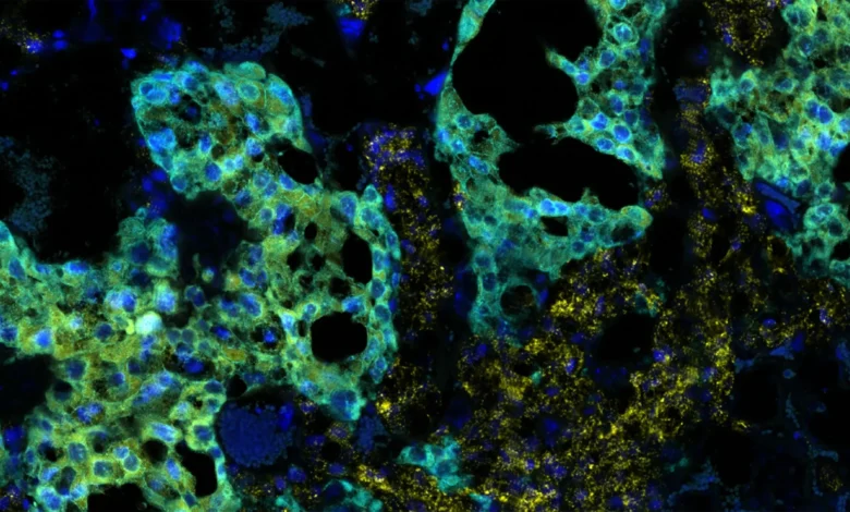
Within this dynamic organ, the researchers focused their attention on a specialized population of cells known as spongiotrophoblasts. These cells occupy a critical interfacial zone within the mouse placenta where maternal vascular networks intimately interface with fetal tissues.
Upon histological examination, a striking dichotomy emerged. In fetuses that continued to develop normally despite the maternal immune challenge, the spongiotrophoblast layer remained structurally intact and healthy. Conversely, male fetuses that subsequently displayed developmental abnormalities exhibited pronounced, visible cellular damage within this specific placental region.
Spongiotrophoblasts perform an indispensable immunological function during pregnancy: they facilitate immune tolerance. Because a fetus inherits genetic material from both the maternal and paternal lines, it is semiallogeneic, meaning it possesses foreign antigens that could theoretically provoke a destructive maternal immune rejection. The placenta actively dampens and manages this immune response to protect the growing organism.
The CSHL study demonstrates that when maternal immune activation triggers severe stress or damage within the spongiotrophoblast layer, this protective immunological shield falters. Once compromised, the localized inflammation and circulating maternal immune signals can breach the placental barrier, directly exposing vulnerable fetal tissues to harmful inflammatory cascades. This discovery repositions the placenta from a passive victim of systemic inflammation to an active participant—and potential gatekeeper—in the pathogenesis of neurodevelopmental divergences.
The Sex Disparity Puzzle: Unlocking the Male Vulnerability
One of the most provocative revelations of the CSHL study is the absolute demarcation of outcomes along biological sex lines. Across every experimental iteration, autism-like behavioral and physiological traits manifested exclusively in male fetuses. Female littermates, exposed to the exact same maternal immune activation and identical environmental conditions, continued their development along typical trajectories without displaying these specific abnormalities.
This sharp divergence mirrors epidemiological trends in human populations, where autism spectrum disorder is diagnosed in males at a rate approximately four times higher than in females. For years, researchers have debated the causes of this pervasive sex bias, proposing theories that range from diagnostic screening disparities and societal behavioral expectations to distinct genetic protective factors.
The CSHL findings inject a powerful biological hypothesis into this ongoing debate by demonstrating that sex-specific vulnerability is established at an extraordinarily early developmental stage.
"It’s really fascinating, because we’re looking at an age which precedes hormonal surges," Cheadle notes, highlighting the timing of the vulnerability window. "It precedes sexual differentiation in the brain and many other regions. So, it’s surprising there would be a sex-based difference this early."
Because the divergence occurs before the classical surges of testosterone and estrogen that drive sexual differentiation in the mammalian brain, researchers must look beyond traditional hormonal explanations. Two primary hypotheses currently guide scientific inquiry into this phenomenon. First, male embryos may express unique antigenic proteins on their cellular surfaces that provoke a more aggressive or structurally damaging response from an inflamed maternal immune system. Second, female embryos may possess intrinsic, proactive cellular defense mechanisms—potentially linked to genetic factors on the X chromosome or distinct metabolic pathways—that shield them from the deleterious effects of inflammation during sensitive developmental epochs.
Cheadle and his team emphasize that unlocking the secret of this female resilience could hold the key to therapeutic innovation. By identifying the molecular mechanisms that naturally protect female fetuses, scientists may be able to engineer pharmaceutical or biological interventions that mimic these protective pathways in vulnerable male pregnancies.
Broader Impact, Industry Implications, and Future Directions
While the current study was conducted entirely using murine models and cannot yet be directly generalized to human clinical diagnoses, its broader implications for maternal-fetal medicine and neurodevelopmental research are substantial. As researchers worldwide strive to construct a more holistic, multi-systemic understanding of autism, studies that integrate immunology, placental biology, and neurodevelopment are rapidly reshaping the scientific landscape.
The realization that critical foundational events in autism-like development may originate within the placenta offers a paradigm shift for early risk assessment. Currently, most evaluations for developmental delays occur postnatally, often years after behavioral symptoms become observable. If future translational research confirms that human placental biomarkers can reflect early gestational immune stress, clinicians might one day be equipped with non-invasive diagnostic tools to identify high-risk pregnancies well before birth.
Furthermore, these insights validate the growing importance of maternal health management during gestation. They reinforce existing clinical guidelines emphasizing the prevention and rapid management of severe viral infections, fevers, and systemic inflammatory conditions during pregnancy. As medical science increasingly recognizes the interconnectedness of maternal immunity and fetal neurodevelopment, prenatal care standards may continue to evolve to incorporate targeted anti-inflammatory strategies for high-risk cohorts.
Looking ahead, the research team at Cold Spring Harbor Laboratory is expanding its investigative scope. Future phases of the project will involve deeper molecular profiling of both male and female placental tissues to isolate the precise signaling pathways responsible for the divergent outcomes. By mapping the exact biochemical crosstalk between the maternal immune system, the damaged spongiotrophoblasts, and the developing fetal brain, scientists hope to discover novel therapeutic targets.
Ultimately, the CSHL findings remind the scientific community that complex conditions like autism cannot be fully comprehended by studying a single organ in isolation. By looking past the confines of the cranium and examining the profound physiological dialogue between mother, placenta, and fetus, researchers are illuminating new pathways toward understanding, diagnosing, and ultimately supporting the diverse spectrum of human neurodevelopment.